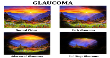
GLAUCOMA
Glaucoma is a group of chronic progressive eye diseases which lead to irreversible damage of the optic nerve situated in the back of the eye. The damage is usually caused by high intraocular pressure (IOP) or pressure inside the eye.
The eye pressure is maintained by a fluid inside the eye also known as AQUEOUS HUMOUR. This fluid inside the eye is drained through fine channels known as Trabecular meshwork. The eye pressure rises if the aqueous is not drained properly or there is excessive production of the aqueous.
The signs or symptoms of glaucoma can vary depending on the type. Primary open-angle glaucoma often develops slowly and painlessly, with no early warning signs. It can gradually destroy one's vision without even knowing it.
The peripheral vision of the vision is first to affected by the nerve damage and if not treated properly it may slowly lead to total blindness. The peripheral vision is involved in the initial phase of glaucoma and patient is completely unaware of the vision loss. Thus, GLAUCOMA is also known as "SILENT THIEF OF VISION"
Acute angle-closure glaucoma results from a sudden blockage of drainage channels in the eye, causing a rapid rise of eye pressure. In this form of the disease, a patient would have blurred vision, the appearance of halos or coloured rings around lights, and pain and redness in the eye.
Glaucoma is one of the leading causes of blindness for people over the age of 60. If not diagnosed and treated properly it can lead to irreversible vision loss and complete blindness.
It can occur at any age but is more common in older adults. After the age of 40 years comprehensive eye examination is absolutely necessary to rule out glaucoma.
Chance of glaucoma increases manifolds in first degree relatives of glaucoma patients. Persons with positive family history should get their comprehensive eye checkup done yearly once.
- The risk of glaucoma rises after the age of 40.
- Family history of glaucoma
- Raised intraocular pressure
- Physical injuries to the eye- Severe trauma, such as being hit in the eye, can result in immediate increased eye pressure.
- Corticosteroid use- Using corticosteroids (including cortisone, hydrocortisone, and prednisone) for prolonged periods of time appears to put some people at risk of getting secondary glaucoma.
- Extremes of refractive errors – like high myopia or high hypermetropia is also a risk factor for glaucoma
- Some studies indicate that diabetes, high blood pressure, and heart disease may increase the risk of developing glaucoma.
- Conditions such as retinal detachment, eye tumours, and eye inflammations may also trigger glaucoma.
Comprehensive eye examination is mandatory for early detection of glaucoma as in most cases there is minimal or no symptoms till advanced stage of glaucoma.
- Patient history to determine any symptoms the patient is experiencing and if there are any general health problems and family history that may be contributing to the problem.
- Visual acuity measurements
- Tonometry to measure the pressure inside the eye
- Pachymetry to measure corneal thickness.
- Gonioscopyto visualize the drainage angle of the eye which helps to determine whether the angle is open or closed.
- Visual field testing, also called perimetry, to check if the field of vision (peripheral and central) has been affected by glaucoma.
- OCT (Optical Coherence Tomography): This is a test to see the status of the optic nerve head and the thickness of the retinal nerve fibre layer within the eye
- Dilated eye examination : In this examination , drops are given to dilate the pupil to visualize the optic nerve and the other structures inside the eye.
There are many types of glaucoma. Some are enumerated here:
- PRIMARY OPEN ANGLE GLAUCOMA
This is the most common form of glaucoma. Damage to the optic nerve is slow and painless. Those affected can lose a large portion of vision before they notice any vision problems.
- ANGLE CLOSURE GLAUCOMA
This type of glaucoma, also called closed-angle glaucoma or narrow-angle glaucoma. It occurs when the drainage angle in the eye (formed by the cornea and the iris) closes or becomes blocked.
The acute form occurs when the iris completely blocks fluid drainage. When people with a narrow drainage angle have their pupils dilated, the angle may close and cause a sudden increase in eye pressure. The initial intraocular pressure is reduced by medical treatment or laser treatment (Peripheral Iridotomy/PI). A laser treatment in the fellow eye is done to prevent the chance of a similar attack.
- NORMAL TENSION GLAUCOMA
In this form of glaucoma, eye pressure remains within the "normal" range, but the optic nerve is damaged nevertheless.
- CONGENITAL GLAUCOMA
Glaucoma can happen at any age although more common in older age group. This type of glaucoma is rare and occurs at birth or soon after. The signs and symptoms include enlargement of the eyes, watering, unusual sensitivity to light or haziness of the anterior transparent portion of the eye called the cornea. Although the initial treatment begins with eyedrops, surgical intervention is mandatory in these cases.
- SECONDARY GLAUCOMA
This type of glaucoma results from an injury or another eye disease. It may be caused by a variety of medical conditions, medications, physical injuries, and eye abnormalities. Other eye surgery can also lead to secondary glaucoma.
IS THERE A TREATMENT OF GLAUCOMA?
There are multiple treatment modalities available for glaucoma. Most of the treatment modalities are aimed to reduce the intraocular pressure and thus slowing the disease progression or halting it.
WHAT ARE THE TREATMENT OPTIONS AVAILABLE?
Glaucoma treatment is aimed at reducing pressure in the eye. Regular use of prescription eye drops are the most common and often the first treatment.
It can be achieved by – 1 medication
2 LASER
3 SURGERY
Glaucoma is characterized by irreversible damage to the optic nerve. The modern treatment amenities does not reverse the damage caused by glaucoma eye hospital in Kolkata, but it can definitely halt or slow down the disease progression by reducing the Intraocular pressure.
The drops and treatments prescribed by the treating ophthalmologist should be followed religiously. During the drop instillation proper care should be taken to prevent wastage. The drops has to be continued lifelong unless and otherwise stopped by the treating ophthalmologists.Adherence to the treatment prescribed is of utmost importance.
Closed angles are opened using YAG laser by making small hole in the peripheral iris (part of eye)- this procedure is also known as YAG peripheral iridotomy or YAG PI.
In open angle cases also high energy laser beams are focused on to the drainage channels to facilitate the outflow of aqueous – this type of laser is known as LASER TRABECULOPLASTY.
The YAG PI procedure is not painful. During the procedure the patient may find slight discomfort which quickly subsides once the procedure is completed. During the procedure a lens is generally placed in front of eye, which may give an uncomfortable feel which is subsided immediately after completion of the procedure.
The procedure is generally done under topical anaesthesia.
One can commence normal activity as soon as half an hour after the completion of procedure. As it is not an intraocular procedure, one does not have to be under any restrictions after the procedure.
One can go to office from very next day. The vision may be blurred on the day of procedure as few medicines are instilled in the eye to facilitate the procedure.
One does not have to wear dark glasses after YAG PI.
There is no such restrictions after completion of YAG PI
The procedure per se takes only 5-10 minutes. Prior to the procedure few medications needs to be instilled in the eyes which may take up around 1-2 hours in total.
- The glaucoma surgeries are indicated if the eye pressure is not controlled with the medical management or the patient is having allergic reactions to multiple antiglaucoma medications.
- The surgical options are as followed:
- TRABECULECTOMY - If eye drops and laser surgery aren't controlling eye pressure, the patient may need a trabeculectomy. This filtering microsurgery creates a drainage flap. Fluid can then percolate into the flap and later drain into the vascular system.
- DRAINAGE IMPLANTS - Drainage valve implant surgery may be an option for uncontrolled Glaucoma. A small silicone tube is inserted in the eye to help drain fluid.
The surgeries can definitely halt or stabilise the disease progression in most of the cases but it can’t reverse or cure the damage that has already occurred.